Optical imaging of chronic pain in the brain
Background
Chronic pain states are accompanied by changes in the brain. Stimuli are perceived more intensely, pain processing is enhanced, and brain activity in the limbic system – the center of emotions – is increased. Thanks to modern imaging techniques, such as functional magnetic resonance imaging (fMRI) and voxel-based morphometry – a three-dimensional quantitative imaging of areas of the brain – brain regions with increased or even reduced activity can be precisely identified; even minimal structural changes are detected.

Research goal
Prof. Paul Geha of the University of Rochester has been studying the morphological and functional differences in the brains of healthy people and patients with chronic pain for many years. Based on his observations, he is certain that the development of chronic pain is anchored here and may even be predictable by imaging. The deeper understanding of the causes of chronic pain and its resolution would open up possibilities to counteract this development with methods of psychotherapy or with specific drug therapy. Areas most likely to respond to analgesic therapy are in the limbic system, where emotions are processed.
Prof. Paul Geha
Paul Geha, MD, is Professor of Psychiatry and Neurology at the University of Rochester in New York State, USA. He is extensively experienced in brain imaging in chronic pain. He was trained in the renowned laboratory of Prof. Vania Apkarian, MD, who postulated in 2008 that the development of chronic pain is an emotional learning process. Together with him, Prof. Geha developed a dynamic concept for the development of chronic pain that involves changes in the circuitry between brain regions.
During his time as an assistant professor at Yale University, he also worked closely with the laboratory of Prof. Stephen Waxman, one of the world leaders in sodium channel research related to pain.
Mercator Fellow for KFO5001
Since there are a number of synergisms between Prof. Paul Geha’s work and the research program of KFO5001, we mutually agreed upon a cooperation. For two months per year, Paul Geha will directly support pain research at the University Hospital of Würzburg. The focus here is on how CNS changes in pain influence the peripheral nervous system. Mercator Fellows are outstanding personalities from science and practice whose involvement in specific research questions is paid for by the German Research Foundation (DFG).
Selected publications
Geha PY, Baliki MN, Harden RN, Bauer WR, Parrish TB, Apkarian AV (2008)
The brain in chronic CRPS pain: abnormal gray-white matter interactions in emotional and autonomic regions.
Neuron. 2008 Nov 26; 60 (4): 570-581
Go to publication
Makary MM, Polosecki P, Cecchi GA, DeAraujo IE, Barron DS, Constable TR, Whang PG, Thomas DA, Mowafi H, Small DM, Geha P (2020)
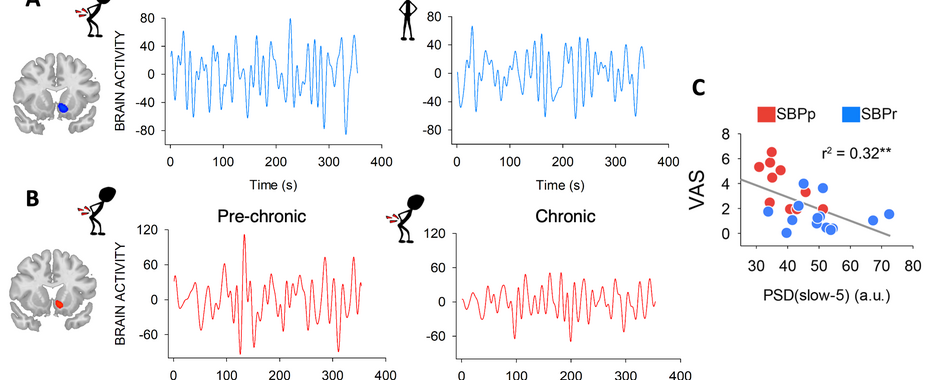
Loss of nucleus accumbens low-frequency fluctuations is a signature of chronic pain.
Proc Natl Acad Sci U S A 2020 May 5;117 (18): 10015-10023
Go to publication
Research Team
Head
Prof. Dr. med. Paul Geha
Mercator Fellow


