
Core Facility Imaging: 7T Whole Body Scanner Magnetom™ Terra
The infrastructure supports human, large animal, and ex-vivo imaging studies, all conducted under established hygienic and safety protocols. The combination of high magnetic field strength, custom RF hardware, and comprehensive physiological monitoring allows researchers to investigate anatomy, function, and molecular processes with high spatial resolution.
The scanner features:
- 3rd-order shimming system for optimal magnetic field homogeneity
- 8-channel RF Power Amplifier (RFPA) supporting parallel transmit (pTX) technology
- A range of commercial and home-built specialized RF coils with pTX support for different applications
Human Imaging
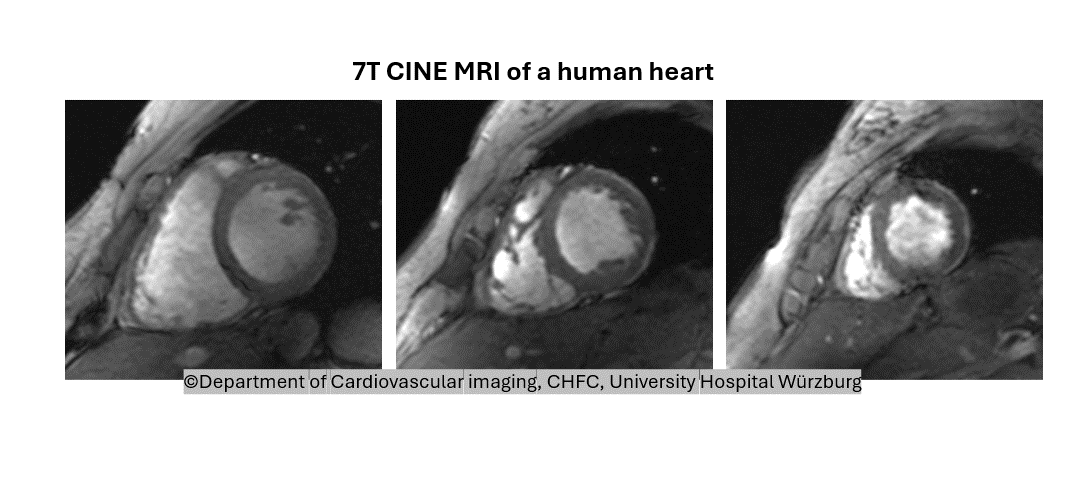
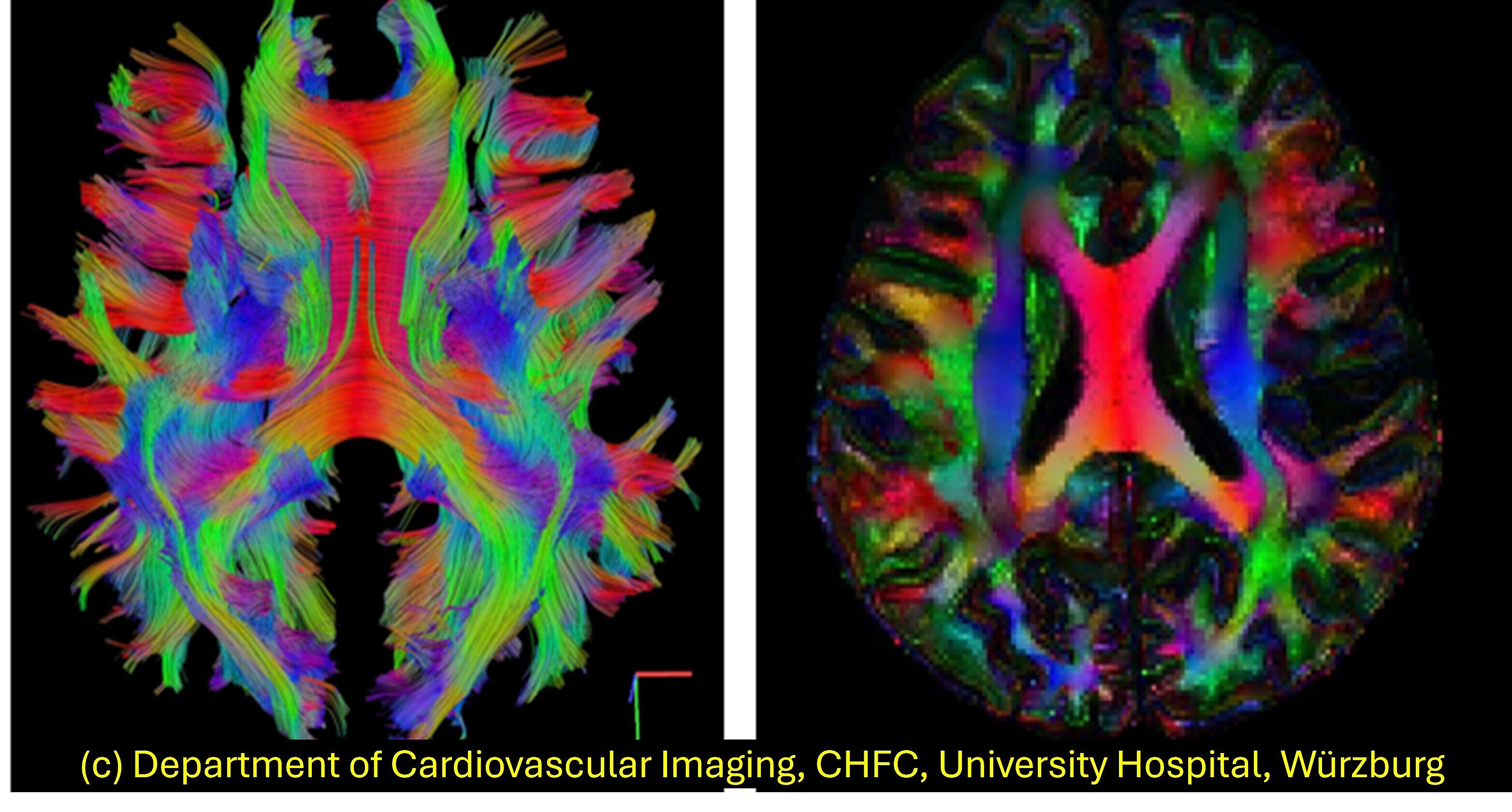
Human MRI studies are conducted using dedicated commercial coils for the head and thorax, with strict adherence to hygienic standards. Cardiac MRI capabilities include dynamic CINE imaging for detailed visualization of cardiac motion and function, and T2*-weighted imaging for myocardial tissue characterization, especially useful in detecting iron deposition or hemorrhage. A custom-built cardiac RF coil featuring 8 transmit and 16 receive channels is available to support parallel transmit sequences, enhancing image quality and reducing artifacts. Brain MRI is another major focus and includes high-resolution structural scans (including DTI) and functional MRI (fMRI) for psychiatric and cognitive research. fMRI studies are conducted in cooperation with the Department of Psychiatry, which provides access to specialized expertise and equipment, enabling advanced BOLD imaging and brain activation mapping in various experimental protocols.

7T Diffusion Tensor Imaging of Human Brain
Animal Imaging
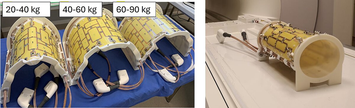
The facility also supports advanced imaging of large animals, with a primary focus on cardiac MRI in pigs for studying myocardial infarction and cardiovascular remodeling. The imaging system is integrated with a nearby large animal research facility and includes a dedicated preparation room, enabling streamlined workflows and adherence to animal welfare regulations. Custom in-house developed pTX RF coils are available for pigs ranging in weight from 20 to 90 kg[2-4], allowing adaptation to individual subjects and enabling longitudinal studies. This capability facilitates repeated, high-resolution cardiac imaging to monitor disease progression or therapeutic response over time. In addition to proton imaging, the scanner is equipped to perform X-nuclei imaging, broadening the range of biological processes that can be studied. A dedicated 19F RF coil enables non-invasive tracking of fluorine-labeled immune cells, allowing the visualization of inflammatory responses in vivo. This molecular imaging capability enhances translational research by supporting real-time monitoring of immune cell migration and inflammation in cardiovascular and other disease models.

Ex-vivo Imaging
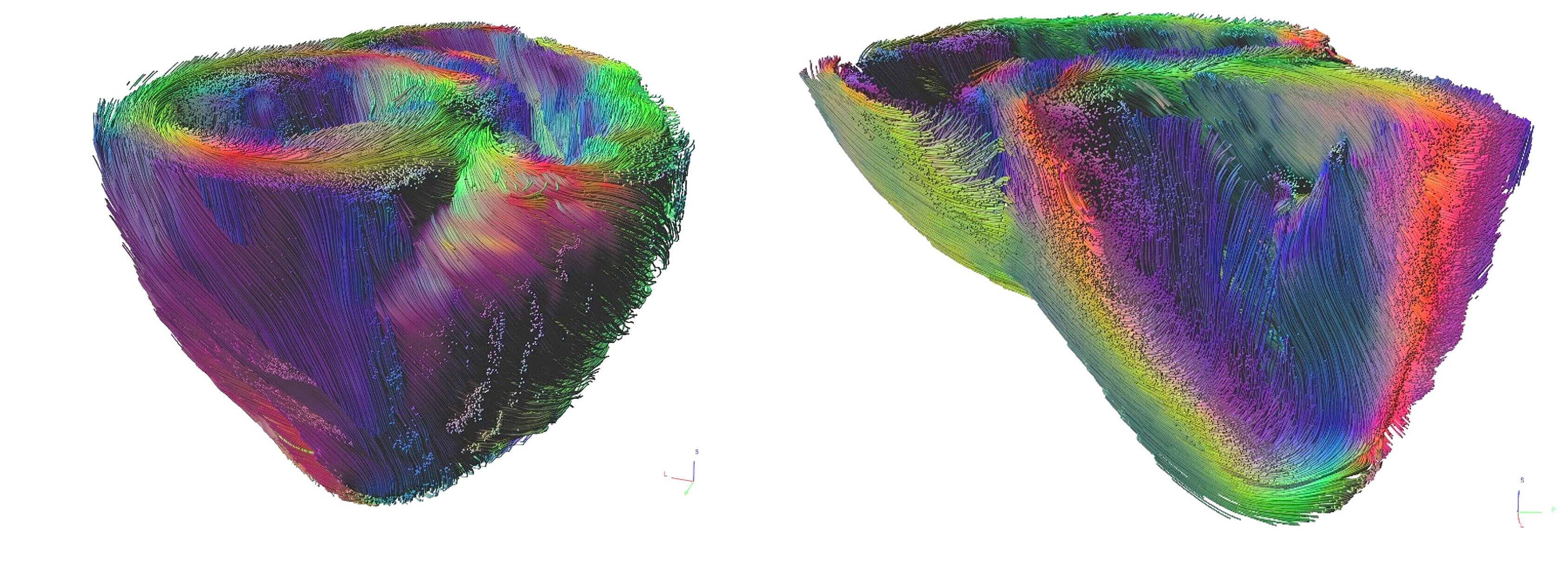
Ex-vivo imaging is an essential component of the facility, allowing ultra-high-resolution scanning of excised and fixed biological samples such as hearts and brains. The 7T scanner's powerful magnetic field and long scan time capabilities enable detailed visualization of anatomical structures at microscopic resolution, free from motion artifacts. Both standard and custom-designed small-diameter RF coils[5] are employed to optimize image quality for varying specimen sizes and geometries. Anatomical imaging protocols reveal fine structural details, while diffusion tensor imaging (DTI) provides insights into tissue microstructure, including fiber orientation and integrity. These ex-vivo imaging capabilities are critical for validating in vivo findings, conducting correlative studies with histology, and developing detailed anatomical atlases in both preclinical and clinical research contexts.
Ex-vivo Diffusion Tensor Imaging of fixed pig heart
Available Additional Equipment
To support a wide range of experimental requirements, the facility is equipped with extensive MRI-compatible auxiliary systems. These include a ventilation machine for respiratory support, a perfusion system for fluid delivery, and an angiography pump for precise contrast agent injection. A fully integrated physiological monitoring system is available, offering real-time ECG, non-invasive blood pressure, pulse oximetry, CO₂ analysis, and temperature measurement using an optical PhotoTemp thermometer. Dedicated RF filter plates allow the safe transmission of electrical signals into and out of the Faraday cage surrounding the MRI suite, facilitating the integration of external devices without compromising image quality or safety. Additionally, wall-mounted safety rails have been installed throughout the scanner room to securely fix equipment with limited MRI compatibility, such as components containing minor ferromagnetic materials. These safety features ensure the secure and stable positioning of auxiliary devices during scanning procedures and mitigate risks in the high magnetic field environment.
Bibliography
- Schreiber, L.M., et al., Ultra-high field cardiac MRI in large animals and humans for translational cardiovascular research. Front Cardiovasc Med, 2023. 10: p. 1068390.
- Elabyad, I.A., et al., A Novel Mono-surface Antisymmetric 8Tx/16Rx Coil Array for Parallel Transmit Cardiac MRI in Pigs at 7T. Sci Rep, 2020. 10(1): p. 3117.
- Elabyad, I.A., Terekhov, M., Bille, M., Schreiber, L.M., Design and Implementation of Two 16-Element Antisymmetric Transceiver Coil Arrays for Parallel Transmission Human Cardiac MRI at 7 T. IEEE Transactions on Microwave Theory and Techniques, 2021. 69(7): p. 3540-3557.
- Elabyad, I.A., et al., A novel antisymmetric 16-element transceiver dipole antenna array for parallel transmit cardiac MRI in pigs at 7 T. NMR Biomed, 2022. 35(8): p. e4726.
- Terekhov, M., et al., High-resolution imaging of the excised porcine heart at a whole-body 7 T MRI system using an 8Tx/16Rx pTx coil. MAGMA, 2023. 36(2): p. 279-293.