
Longitudinal Ultra-High Field cardiac MRI studies of Myocardial Infarction in a Porcine Model
Development of Dedicated RF Coils for Pigs with Parallel Transmit Technology
To address the unique challenges of imaging at ultra-high field strengths, we have designed and implemented custom radiofrequency (RF) coil arrays tailored for pig anatomy [2]. These coils support parallel transmit (pTx) technology, which enables spatially tailored RF excitation to optimize signal-to-noise ratio (SNR) and image contrast—particularly important in sequences involving saturation and inversion recovery pulses, such as perfusion imaging and late gadolinium enhancement.

CINE MRI for Cardiac Function Assessment

We employ high spatial and temporal resolution CINE MRI to non-invasively quantify cardiac function. This technique allows for precise measurement of ventricular volumes, ejection fraction, and wall motion abnormalities, providing critical insights into the impact of myocardial infarction on heart performance.[3]
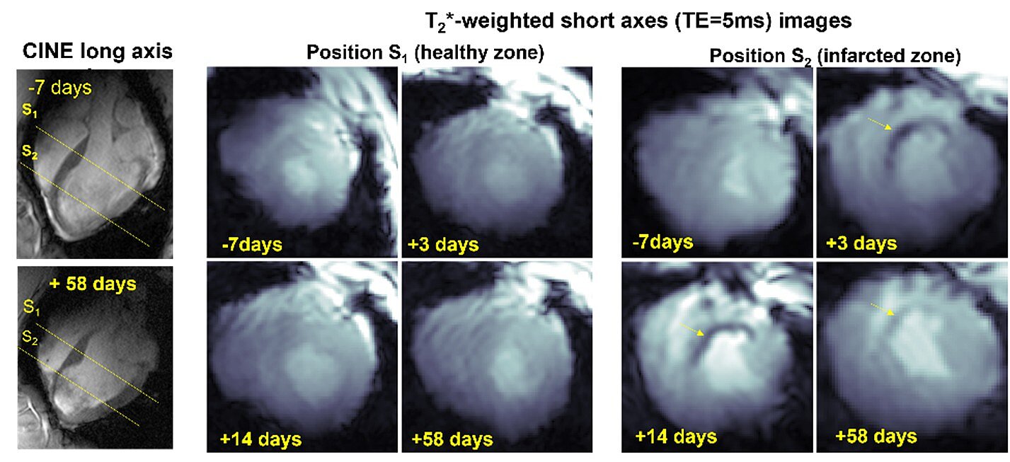
T2* weighted imaging of Post-Infarction Hemorrhage
T2*-weighted imaging at ultra-high field enables sensitive detection and quantification of intramyocardial hemorrhage - a marker of severe reperfusion injury. Our protocols provide high-resolution T2* maps that help characterize tissue integrity and predict adverse remodeling.[4]
First-Pass Perfusion MRI with Gadolinium-Based Contrast Agents

We perform dynamic contrast-enhanced first-pass perfusion MRI to evaluate myocardial blood flow and detect regions of ischemia. This technique offers high temporal resolution imaging during the initial passage of gadolinium contrast, aiding in the identification of perfusion deficits immediately after infarction.[1]
Characterization of Infarction Using Late Gadolinium Enhancement (LGE)
Late gadolinium enhancement imaging is employed to delineate the extent of myocardial infarction and fibrosis. At ultra-high field. LGE provides superior contrast and spatial resolution, facilitating precise quantification of infarct size and viable myocardium.
Late Gadolinium Enhancement of infarcted pig heart

References
- Schreiber, L.M., et al., Ultra-high field cardiac MRI in large animals and humans for translational cardiovascular research. Front Cardiovasc Med, 2023. 10: p. 1068390.
- Elabyad, I.A., et al., A Novel Mono-surface Antisymmetric 8Tx/16Rx Coil Array for Parallel Transmit Cardiac MRI in Pigs at 7T. Sci Rep, 2020. 10(1): p. 3117.
- Lohr, D., et al., Precision imaging of cardiac function and scar size in acute and chronic porcine myocardial infarction using ultrahigh-field MRI. Commun Med (Lond), 2024. 4(1): p. 146.
- Terekhov M., L.D., Hock M., Bille M., Baltes S., Elabyad I., Schnitter F., Aures J., Reiter T., Bauer W., Hofmann U., Schreiber L. Visualization of Post-Infarction Cardiac Tissue Remodeling at 7T using T2* Methodology : Longitudinal Pilot Study in a Porcine Myocardial Infarction Model. in International Society of Magnetic Resonance in Medicne, Annual Meeting. 2021.