Ultra-High-Field 19F MRI of cardiac inflammation with labeled immune cells
Therefore, the scope of this medical issue is vast, and achieving an accurate diagnosis is often challenging. As a result, therapy is frequently restricted to symptomatic treatment, making it difficult to evaluate the success of the proposed therapies.
Although recent advancements incorporate various imaging techniques such as PET, CT, MRI, optical imaging, and ultrasound imaging, visualizing the inflammatory process remains a significant challenge, primarily because the affected tissue does not exhibit specific characteristics in the initial phase that could help differentiate between inflamed and healthy regions. To address this, we are utilizing 19F as a contrast agent through nanoparticles containing perfluorocarbons (PFCs). Additionally, we have access to a 7T MRI Siemens scanner, which we use to noninvasively examine the organs (heart, vessels, liver, lung, bone marrow) of large animals (pigs) using 1H/19F magnetic resonance imaging (MRI). While the magnetic field strength of a clinical MRI unit typically ranges from 1.5 to 3 Tesla, our research unit operates at 7 Tesla.
This requires the design and development of dedicated RF-coils for both 1H and 19F imaging tailored for the pig thorax and capable of overcoming hurdles of MR-imaging at ultra-high magnetic field related to B1+ inhomogeneity.
Design of a dedicated 19F array for 7T MRI in pigs
As shown below, this dynamic setup enables us to capture high-resolution images of the pig heart, providing detailed insights into the morphology of the organ as illustrated below.
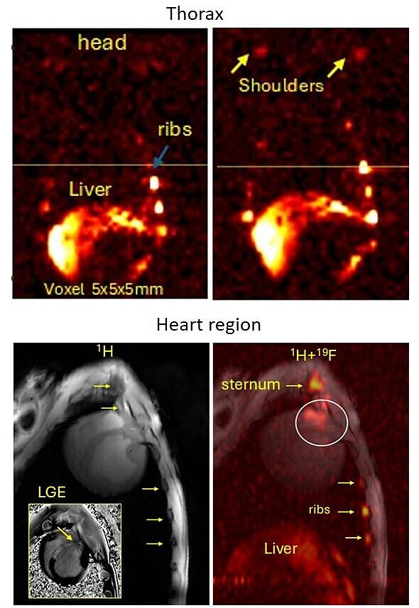
In our current investigations, we are monitoring acute inflammation in the heart following myocardial infarction. The results of this work characterize, for the first time, noninvasive 19F MRI imaging of cardiac inflammation after infarction in a large animal model at 7T MRI, achieved within a reliable short-time acquisition. While translation to the clinic is challenging, it is achievable with the right tools and personnel in place.
In-vivo 7T 19F MRI with labelled immune cells

References
M. Hamid, M. Terekhov, R. Grampp, A. Stadtmüller, M. Keshtkar, I. Elabyad, S. Dembski, G. Ulm, A. Frey, U. Hofmann, W. Bauer, L. Schreiber First Ultra-High-Field 19F MRI in large animal model after Acute Myocardial infarction at 7T MR-scanner, 91-DGK Jahrestagung, https://doi.org/10.1007/s00392-025-02625-4
M. Terekhov, M. Hamid, , R. Grampp, A. Stadtmüller, M. Keshtkar, I. Elabyad, S. Dembski, G. Ulm, A. Frey, U. Hofmann, W. Bauer, L. Schreiber , First 7T 19F MRI with labeled cells in a pig model of myocardial infarction using the twin-rf-array system with pTX support for human MR-scanner, Proceeding ISMRM 2025 # 0478.
M. Hamid, M. Terekhov, R. Grampp, A. Stadtmüller, M. Keshtkar, I. Elabyad, S. Dembski, G. Ulm, A. Frey, U. Hofmann, W. Bauer, L. Schreiber, Optimizing of Flourine-19 at 7T MRI in large animal model after Acute Myocardial infarction. Proceeding ISMRM 2025 # 3070.


