
Functional MRI at 7T: Advancing Brain Research and Artefact Correction
Funded by the Interdisciplinary Center for Clinical Research (IZKF, Project F-461), the project aims to establish 7T neuroimaging in Würzburg and to develop novel methods for the imaging of clinically relevant subcortical structures. Dysfunctions in subcortical regions play a central role in a variety of neuropsychiatric (e.g., anxiety disorders) and neurodegenerative diseases (e.g., Parkinson's), pain disorders, and disorders of central cardiac regulation.
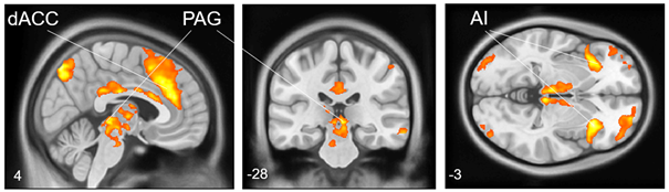
While standard 3T MRI scanners have provided valuable insights into brain function, the achievable resolution is insufficient to visualize small, subcortical brain structures and their subregions. 7T MRI offers significantly higher resolution and sensitivity without temporal penalty in functional monitoring, allowing us to visualize the subcortical fear network in even greater detail.
Project Description
High-resolution imaging of small subcortical regions is challenging, because of susceptibility artifacts that arise in small target regions near large blood vessels or adjacent to ventricles. Moreover, the segmentation of subcortical regions in the human brain is typically poor, which makes a precise anatomical localization of functional activations difficult.
Our research exploits the advantages of 7T MRI to improve functional imaging and addresses these challenges of high field imaging. In more detail, we defined three project areas:
Optimizing protocols for high spatial and temporal resolution functional imaging
Phantom and pilot in vivo measurements were conducted to optimize parameters across various setups. We successfully achieved the target spatial resolution of 1 mm³ isotropic without significantly increasing acquisition time or introducing imaging artifacts. This enables more precise localization of key deep brain regions involved in the fear and anxiety network - structures that are difficult to distinguish on conventional 3T MRI.
Overcoming the challenges of susceptibility artifacts
A major challenge in high-field MRI, particularly at 7T, is image distortion caused by magnetic field inhomogeneities (B₀). These distortions can compromise the accuracy of fMRI, especially in regions with complex tissue boundaries or during acquisitions with long echo times. Our project investigates both prospective online and retrospective offline shimming strategies using a 7T system equipped with advanced third-order shims. These approaches aim to minimize susceptibility artifacts and improve the fidelity of the acquired data. By applying online shimming during participant scanning, we aim to ensure high-quality, distortion-free images while maintaining optimal spatial and temporal resolution in functional brain imaging.
Optimizing structural protocols for high-resolution subcortical regions
To advance our understanding of deep brain structures involved in pain modulation and autonomic regulation, we focus on improving the imaging of the periaqueductal gray (PAG) - a small, deeply located structure surrounded by cerebrospinal fluid spaces. Its size and anatomical complexity pose challenges for conventional MRI. To enhance the visibility and delineation of the PAG and similar regions, we employ ultra-high-resolution structural imaging with carefully tuned sequence parameters (e.g., spatial resolution, contrast weighting, and echo time). These refinements improve contrast-to-noise ratio and anatomical precision, facilitating reliable segmentation and quantification of subcortical nuclei and their subregions.
With this project, we aim to set new standards in ultra-high field functional brain imaging in Würzburg and provide researchers with powerful, off-the-shelf tools to investigate their research questions with unprecedented precision.
Principle Investigators
Prof. Grit Hein
Department of Psychiatry, Psychosomatics, and Psychotherapy, University of Würzburg, Würzburg, Germany
Prof. Matthias Gamer
Department of Psychology, University of Würzburg, Würzburg, Germany
Dr. Maxim Terekhov
Molecular and Cellular Imaging, Comprehensive Heart Failure Center, University Hospital Würzburg, Würzburg, Germany
Collaboration partners
Prof. Martin Herrmann
Department of Psychiatry, Psychosomatics, and Psychotherapy, University of Würzburg, Würzburg, Germany
Dr. Anna Linda Leutritz
Department of Psychiatry, Psychosomatics, and Psychotherapy, University of Würzburg, Würzburg, Germany
Ebru Ecem Tavacioglu
Department of Psychology, University of Würzburg, Würzburg, Germany
Jasper Bischofberger
Department of Psychiatry, Psychosomatics, and Psychotherapy, University of Würzburg, Würzburg, Germany
Istvan Homolya
Molecular and Cellular Imaging, Comprehensive Heart Failure Center, University Hospital Würzburg, Würzburg, Germany