
Core Facility Imaging: Preclinical MRI of Small Animals
The core facility primarily supports cardiac MRI in mice, providing CINE imaging sequences to evaluate cardiac function with precision. CINE imaging allows the generation of dynamic heart images throughout the cardiac cycle, enabling the assessment of parameters such as ejection fraction, stroke volume, and wall motion. This service is particularly valuable for researchers studying cardiovascular diseases, heart failure, and cardiac remodeling in mouse models [1].
Advanced molecular imaging
In addition to conventional anatomical and functional cardiac imaging, the facility supports advanced molecular imaging using ¹⁹F MRI, enabling non-invasive tracking of cardiac inflammation through fluorine-labeled agents. This technique is ideal for studies investigating myocarditis, immune responses, and the effects of immunomodulatory therapies. The scanner also supports ²³Na MRI, allowing researchers to explore sodium concentration changes in the mouse body or rat brain, which is relevant in stroke, tumor, or metabolic studies. Both in vivo and ex vivo imaging protocols can be customized based on research needs, including brain, heart, abdominal, and musculoskeletal imaging.
Multi-nuclear imaging
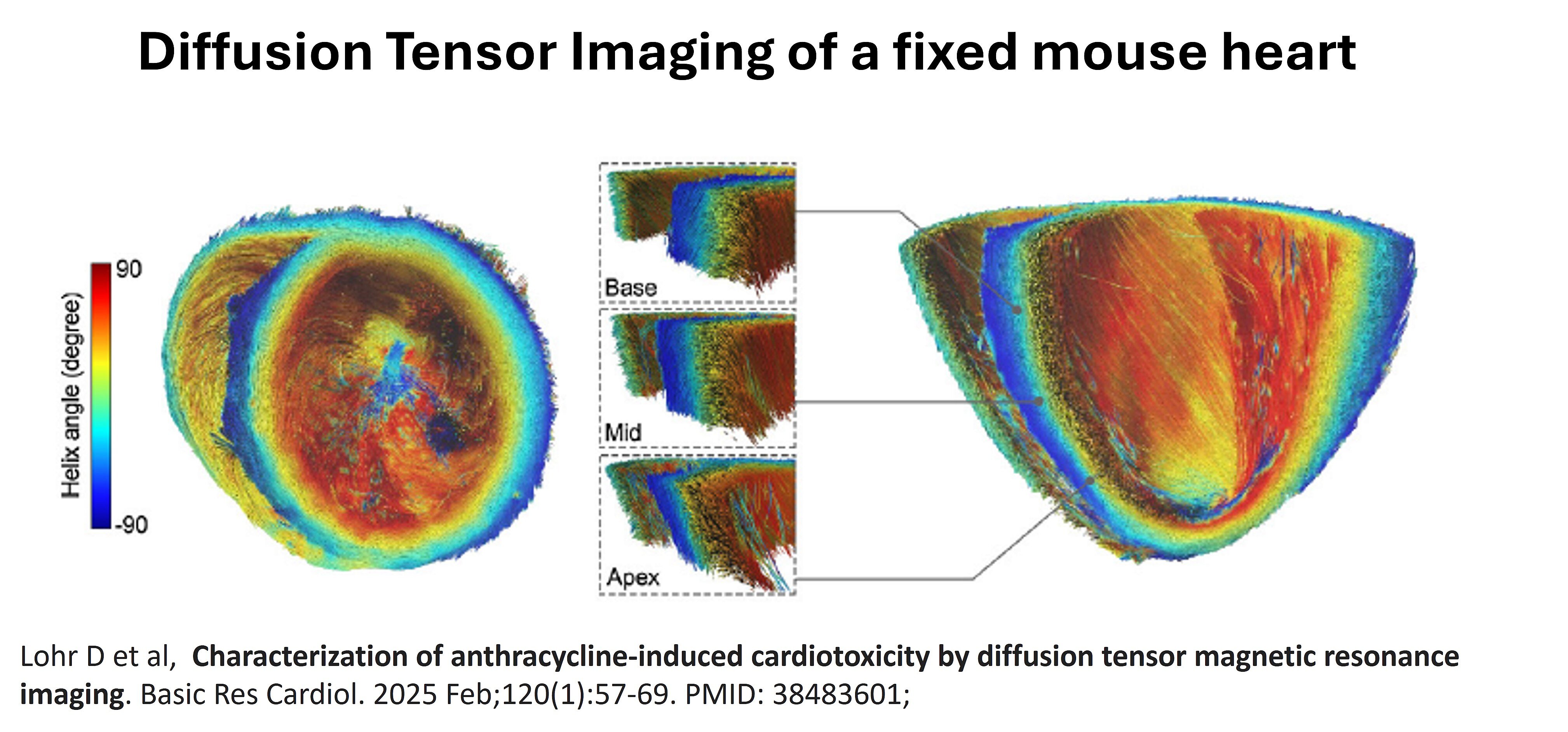
The service supports multi-nuclear imaging, extending applications beyond ¹H to nuclei such as ¹⁹F and ²³Na, enhancing research in metabolic, neurological, and inflammatory processes. Researchers can also access diffusion-weighted imaging (DWI) and diffusion tensor imaging (DTI) for microstructural tissue assessment[2].
Preclinical MRI infrastructure
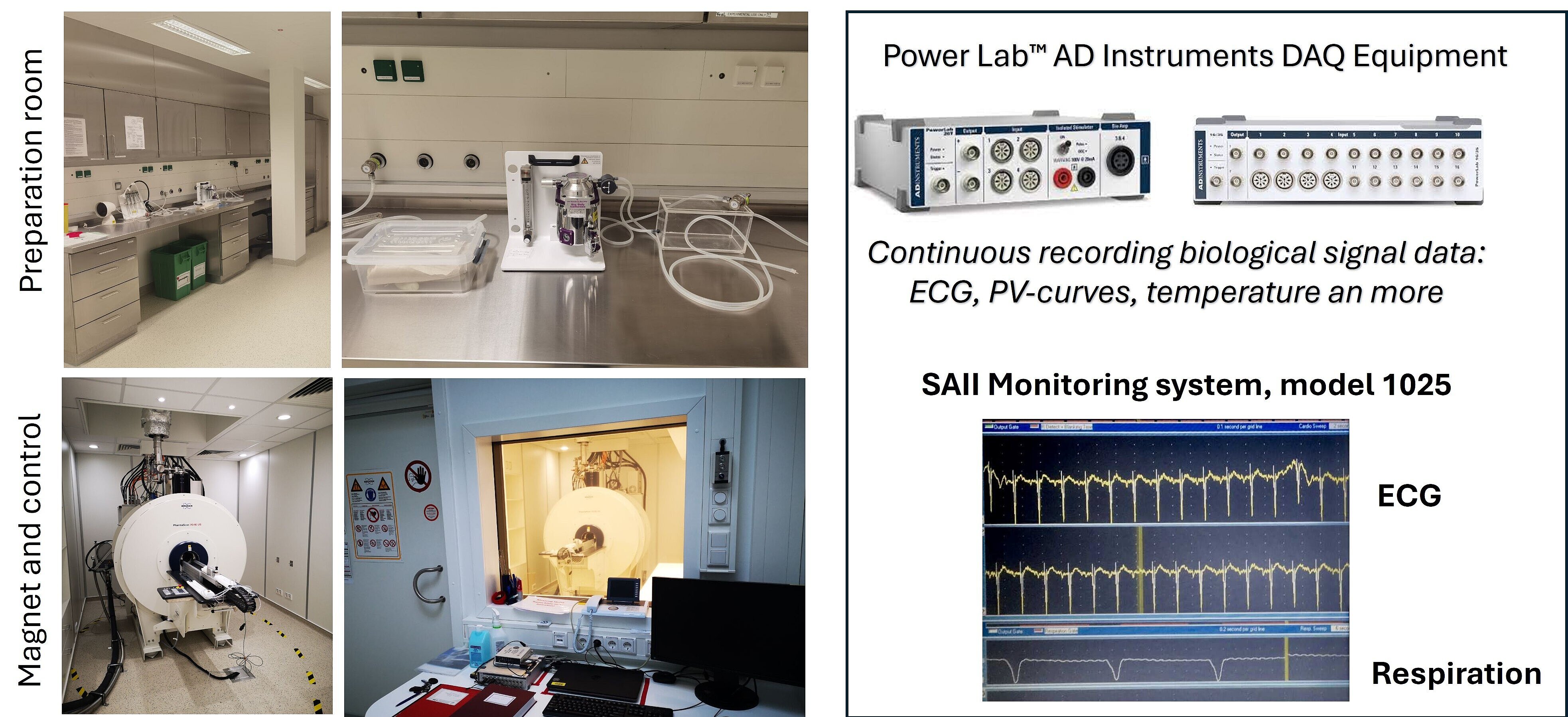
The scanner is equipped with specialized radiofrequency coils, both commercial and in-house designed, including cryogenic coils optimized for mouse and rat body parts, ensuring high-quality data acquisition.
Animal monitoring systems integrated with the MRI allow for precise control of temperature, respiration, and cardiac gating, which is essential for longitudinal and functional studies. The facility is staffed by experienced imaging scientists who assist with protocol development, data acquisition, and analysis. Post-processing support includes advanced cardiac analysis, image registration, segmentation, and quantitative mapping.
Tracking of disease progression or therapeutic response
In addition to service provision, the core supports method development and pilot studies, helping users tailor imaging protocols for novel applications. Users can perform longitudinal studies, allowing the tracking of disease progression or therapeutic response over time. Ex-vivo imaging options include high-resolution anatomical scans, useful for correlating histology and MRI.
Connection with Clinical Imaging Core Facility allows seamless translational research
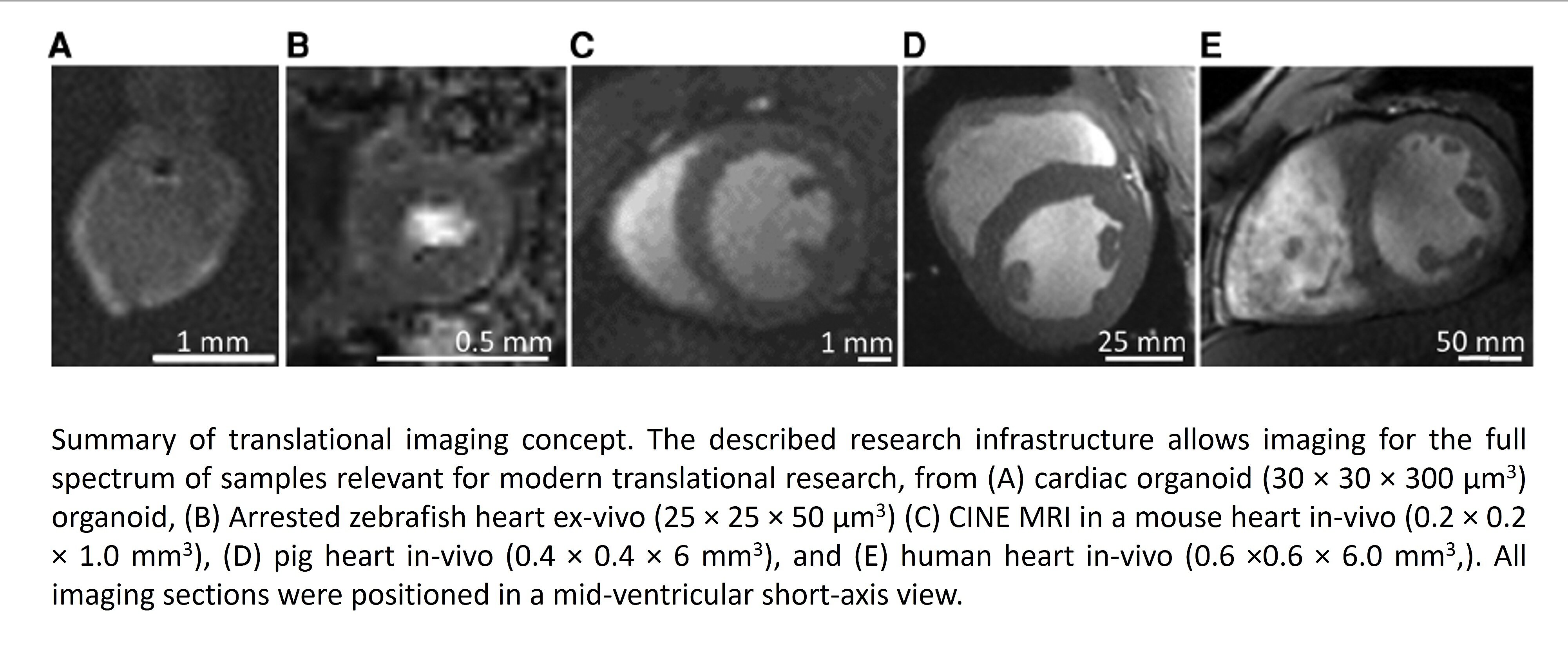
Data outputs are provided in standard formats, compatible with commonly used image analysis software. Researchers are guided through ethical and regulatory aspects of animal imaging and MRI safety. The facility fosters interdisciplinary collaboration and encourages the integration of MRI data with other modalities such as PET or optical imaging. The Preclinical Imaging Core Facility is closely integrated with the Clinical Imaging Core Facility, which operates a whole-body 7T MRI scanner for human and large animal studies. This integration enables seamless translational research, allowing direct comparison and validation of imaging biomarkers and protocols across small animal, large animal, and human subjects. Researchers can design studies that progress from preclinical mouse models to large animal validation and ultimately to clinical application, using consistent imaging modalities and field strengths.
Bibliography
- Schreiber, L.M., et al., Ultra-high field cardiac MRI in large animals and humans for translational cardiovascular research. Front Cardiovasc Med, 2023. 10: p. 1068390.
- Lohr, D., et al., Characterization of anthracycline-induced cardiotoxicity by diffusion tensor magnetic resonance imaging. Basic Res Cardiol, 2025. 120(1): p. 57-69.