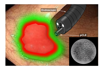
KLEVER- Confocal laser endomicroscopy for real-time visual detection of resection borders
In critical anatomical regions, this may lead to severe consequences such as permanent stomas. Confocal laser endomicroscopy (CLE) has emerged as a promising non-invasive imaging modality, enabling real-time, in vivo visualization of epithelial structures at near-cellular resolution and offering the potential for an “optical biopsy” without tissue removal.
This project aims to develop algorithms for the automated analysis of CLE images to distinguish malignant from benign tissue based on histopathological annotations.
The AI-generated results will be visualized in real time and integrated into conventional white-light endoscopy through an intuitive, user-centered interface. By projecting tumor boundaries directly into the surgeon’s field of view, the system aims to support more accurate intraoperative decision-making and enable precise, organ-preserving resections.
The project is developed in collaboration with Priv.-Doz. Dr. med. Miguel Goncalves (Project lead) and Priv.-Doz. Dr. med. Sven Flemming from University Hospital Würzburg, Prof. Dr.-Ing. Katharina Breininger (CAIDAS, Julius Maximilians University of Würzburg) and KITE Design Research GmbH.


